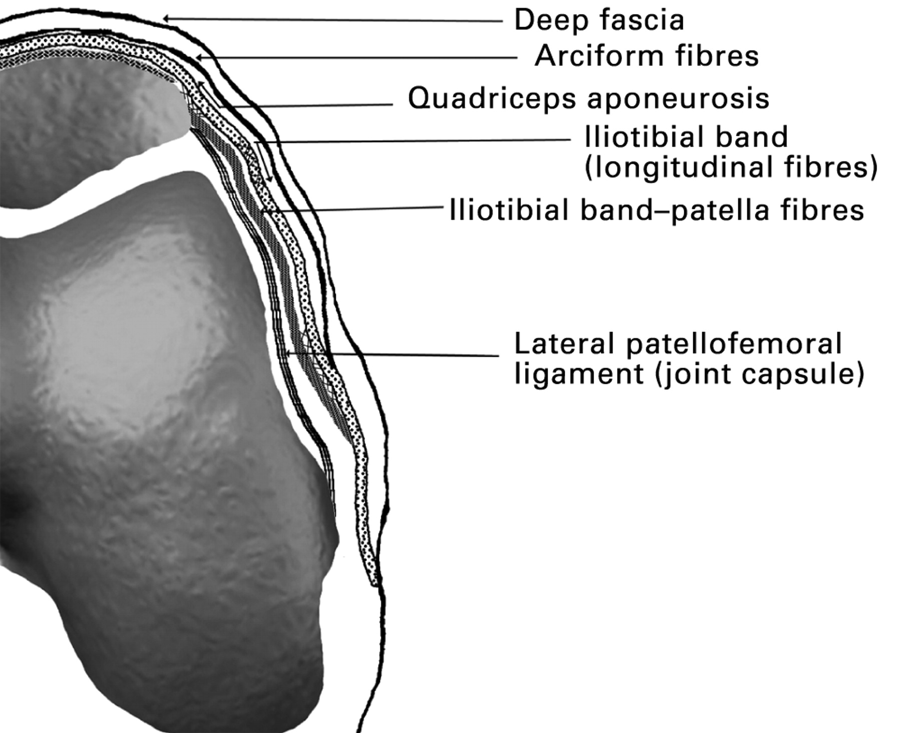
Anatomical Overview
The medial retinaculum, a thick fibrous band, is a crucial structure within the wrist. It serves as a strong and supportive ligament that stretches across the carpal bones, forming the carpal tunnel’s floor. This tunnel, located on the palmar side of the wrist, provides a passageway for tendons and the median nerve.
The medial retinaculum, a fibrous band that helps stabilize the wrist joint, plays a crucial role in hand function. Its intricate interplay with other structures mirrors the dynamic nature of the Mark Cuban Mavericks sale , where players come and go, but the team’s spirit remains unyielding.
Like the medial retinaculum, it serves as a constant amidst the ever-changing landscape of sports, anchoring the team’s foundation and guiding its path towards success.
Location and Structure
The medial retinaculum originates from the pisiform bone and the hook of the hamate bone. It then courses obliquely across the carpal bones, attaching to the scaphoid and trapezium bones. This ligament’s structure resembles a strong, fibrous sheet that covers the carpal bones’ palmar surface, creating a protective roof for the tendons and median nerve.
Medial retinaculum, the thick fibrous band that holds tendons in place, plays a crucial role in hand movement. Like the rhythmic dance steps of Theresa Randle in theresa randle bad boys 4 , it ensures smooth and controlled motion. Just as the medial retinaculum stabilizes tendons, it provides a foundation for graceful movement, allowing us to effortlessly perform everyday tasks.
Relationship with Surrounding Landmarks
The medial retinaculum’s close proximity to various anatomical landmarks plays a significant role in its function. It lies adjacent to the carpal bones, forming the carpal tunnel’s floor. This tunnel houses the flexor tendons of the fingers, as well as the median nerve, which provides sensation to the thumb, index, middle, and ring fingers.
The medial retinaculum, a ligament in the wrist, plays a crucial role in stabilizing the carpal bones. Similar to the intensity of a Clemson player’s ejection , the medial retinaculum ensures the wrist’s stability during strenuous activities, preventing dislocations and ensuring optimal hand function.
Functional Role: Medial Retinaculum

The medial retinaculum plays a crucial role in stabilizing the tendons and supporting the wrist joint. Its primary function is to prevent the tendons from dislocating and ensure smooth movement during wrist flexion.
Preventing Bowstringing of Flexor Tendons, Medial retinaculum
The medial retinaculum is essential in preventing bowstringing of the flexor tendons. Bowstringing occurs when the tendons bulge out or protrude from the wrist crease during flexion. This condition can lead to pain, discomfort, and impaired hand function.
The medial retinaculum acts as a protective barrier that holds the tendons in place and prevents them from bulging outward. By maintaining the tendons’ alignment, the retinaculum ensures efficient and pain-free wrist flexion.
Clinical Significance

The medial retinaculum plays a crucial role in protecting the tendons and nerves within the carpal tunnel. However, injuries to this structure can lead to debilitating conditions that affect hand function.
The most common injury associated with the medial retinaculum is carpal tunnel syndrome. This condition arises when the retinaculum becomes thickened and inflamed, compressing the median nerve as it passes through the carpal tunnel.
Surgical Procedures
In cases of severe carpal tunnel syndrome, surgical intervention may be necessary to release the pressure on the median nerve. The most common surgical procedure is called carpal tunnel release.
- During carpal tunnel release, the surgeon makes a small incision in the palm and divides the medial retinaculum, creating more space for the median nerve.
- This procedure effectively relieves pressure on the nerve, alleviating symptoms such as numbness, tingling, and pain.
Rehabilitation and Exercises
After carpal tunnel release surgery, patients undergo a rehabilitation process to restore wrist function and strength.
- Rehabilitation typically involves a combination of exercises, including range-of-motion exercises, strengthening exercises, and nerve gliding exercises.
- These exercises help to improve flexibility, strengthen the wrist muscles, and promote nerve recovery.
The medial retinaculum, a fibrous band that stabilizes the flexor tendons in the wrist, shares a surprising connection with miriam adelson israel. As a philanthropist and advocate for Israel, Adelson’s unwavering support mirrors the medial retinaculum’s unwavering role in maintaining wrist stability.
The medial retinaculum, a crucial ligament in the wrist, plays a vital role in stabilizing the carpal bones. Its intricate structure, like the symphony of a maestro, ensures seamless movement and dexterity. Interestingly, the renowned quarterback Patrick Mahomes relies heavily on the stability provided by his medial retinaculum to unleash his extraordinary throws, showcasing the profound connection between the human body and athletic prowess.